Patellar tendinopathy, also known as jumper's knee, is a common overuse injury that affects the patellar tendon, which connects the kneecap (patella) to the front of the shinbone (tibia). This tendon is responsible for transmitting force and load from the quadriceps muscles in the front of the thigh to the shinbone, allowing the knee to extend (straighten). This is required for walking, running and in particular, jumping.
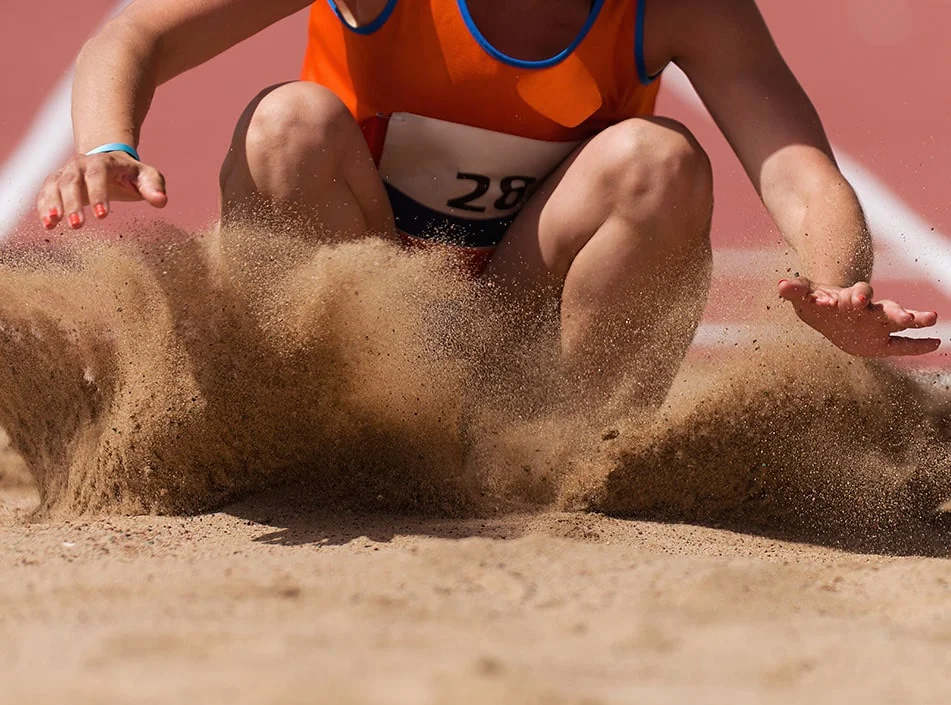
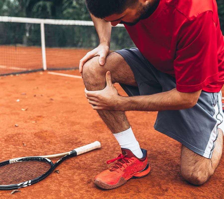
Patellar tendinopathy is most common in athletes who participate in activities that involve repetitive jumping, running, and changing directions, such as basketball, volleyball, soccer, and tennis. It can also occur in individuals who engage in high-impact activities, such as running or aerobics, particularly as a result of sudden changes in their training program.
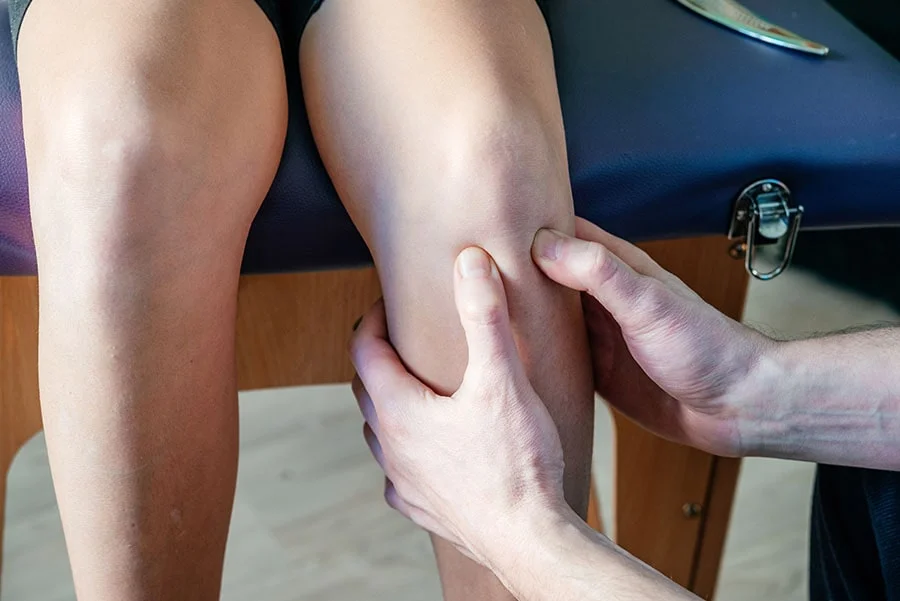
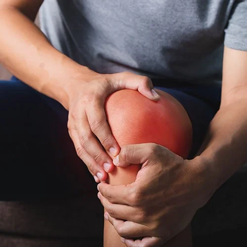
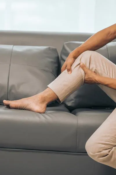
The main symptom of patellar tendinopathy is pain in the front of the knee, typically around the bottom of the kneecap, at the upper end of the tendon. The pain is often aggravated by activities that involve jumping, running, or when walking down stairs. Other symptoms may include:

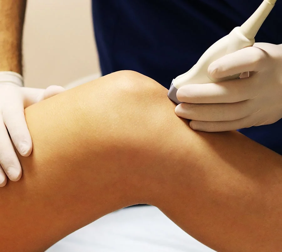
A doctor will typically diagnose patellar tendinopathy based on a medical history, physical examination, and imaging tests. The physical examination will focus on assessing for localised tenderness and pain at the top end of the tendon, as well as range of motion in the knee. Imaging tests, such as an ultrasound can be a very helpful way of confirming the diagnosis and ruling out other potential causes of knee pain. On ultrasound assessment your doctor will look for thickening of the tendon. The maximum normal tendon diameter in ~5.5mm from the most superficial part to the deepest part. In patellar tendinopathy the tendon can become thickening and swollen, sometimes to four times the normal diameter (~20mm). A tendinopathic tendon will also look structurally different on ultrasound with a mixed picture of both darker and lighter areas of the tendon, usually found in the central part of the tendon, at the top end, in the deep part. The clinician will also assess for an increase in blood flow within the tendon, which is not normally seen in a healthy tendon but can be seen floridly with patellar tendinopathy.
The treatment for patellar tendinopathy typically involves conservative measures, such as:
In some cases, additional treatment options may be considered, such as:
At The Joint Injection Clinic, these injections are performed after a thorough consent process, whereby the risk and benefits of the procedure are discussed in detail with your doctor. The experienced medical doctor will then place you in a lying position, face up. The skin is cleaned using a cleaning solution to ensure that the procedure is performed under sterile conditions. Local anaesthetic is injected from the skin to the tendon under ultrasound guidance. After giving the local anaesthetic a few minutes to take effect, the interface between the tendon and the Hoffa’s fat pad is injected with local anaesthetic, water or saline for injection and occasionally, a small dose of steroid.
The high volume injection itself is normally completed within two to three minutes, after which a plaster is applied and post-injection advice is given. The patient is advised to look out for any signs of infection, specifically to check whether the local area becomes red, hot, tender, swollen or if they develop a fever. If this occurs then the patient is asked to contact the clinic immediately at which time a formal reassessment will occur and if needed oral antibiotics can be prescribed. The patient is also warned that following any injection they may notice a short-term worsening or flare in their symptoms after the local anaesthetic has worn off (4-5 hours). This may last for 3-5 days and the patient is advised to consider icing of the area using an ice pack for 10-15 minutes every hour as required.
Surgery is rarely considered for patellar tendinopathy and is typically only recommended if conservative measures have failed to provide relief.
There are several things you can do to help prevent patellar tendinopathy, such as:
